


Dermatophytes and dermatophytosis in humans and animals

Recognition and treatment of dermatophytosis


It is important to know how to identify a dermatophyte.
Then it's easier to heal it too.
It is extremely important to know what type of dermatophyte it is. If we recognize the type of dermatophyte, we can also assume the source of the infection. And if we know the source of the infection, we can try to prevent new infections of animals and humans.
Dermatophytes can be identified by their morphological characteristics, which are divided into two stages: parasitic and saprophytic stage.
Parasitic stage of dermatophytes
In the parasitic stage, a dermatophyte is present in the form of spores (so-called arthrospores) as a sheath around the hair or in the hair itself. The arthrospores can be seen under the microscope.
There are slight differences between arthrospores of different types of dermatophytes, but they are not easy to spot. Therefore, if we find dermatophyte arthrospores, we can be sure that it is dermatophytosis.
More about arthrospores see in video file.
Source - author: youtube.com - Microhub Plus


Dermatophyte arthrospores can be seen in various laboratory procedures. It is important to notice arthrospores around or in the hair, depending on the type of dermatophyte. The presence of arthrospores indicates dermatophytosis but not the type of dermatophyte. This requires a further laboratory procedure of cultivation on mycological artificial nutrient media.
Saprophytic stage of dermatophytes
In order to find out the exact type of dermatophyte, we need to use more complex methods of diagnostics, i.e. recognition of morphological forms in the so-called saprophytic stage of dermatophytes.
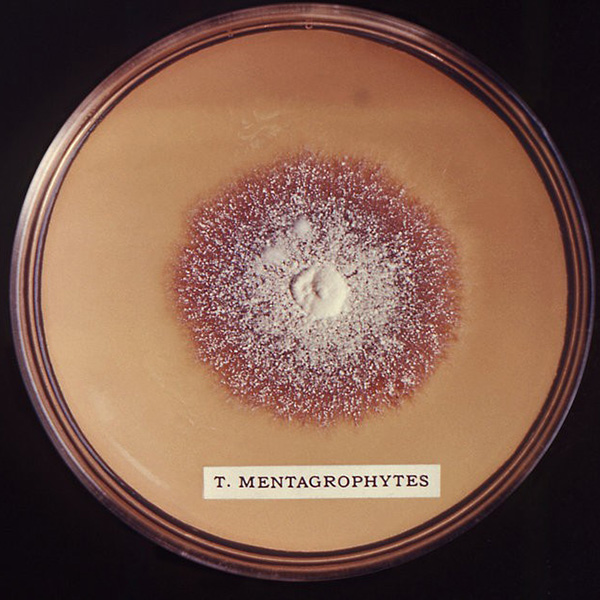
The characteristic dermatophyte formations in the saprophytic stage is produced on artificial nutrient media, which we use in a mycological laboratory, a place that is equipped to work and identify different types of fungi.

Dermatophyte laboratory-cultivated on artificial nutrient media.

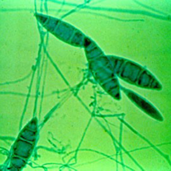
This is what the dermatophyte Microsporum canis looks like. It was extracted from cat and dog fur where we noticed bald areas.

Macroconidia: spindle-shaped multicellular formations and mycelium, accumulation of hyphae - long cells found in fungi.

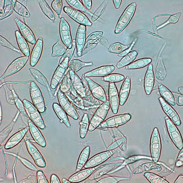
This is what the dermatophyte Trichophyton mentagrophytes looks like. It was extracted from hamster fur where we noticed various bald areas.

Description of macroconidia: cigar-shaped multicellular formations and convoluted hyphae - elongated cells.


Treatment of dermatophytosis

Dermatophytoses in humans
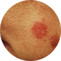
In humans, healthcare professionals make a diagnosis of dermatophytosis. If we notice any suspected skin change caused by a dermatophyte, we should seek medical attention. It is good to remember where we had spent time before the skin changes occurred and what animals we had been in contact with. From our story, the healthcare professional will try to determine the possible source of infection and may advise us to take our pet to the veterinarian.
After the examinations and depending on the clinical changes in the person, the healthcare professional determines the treatment. The basis for treating dermatophyte changes is the use of antifungal drugs called antimycotics. The form of administration of these drugs may vary depending on the severity of the skin changes. The most important thing is to follow exactly the instructions that each healthcare professional will give to his patient in order to recover as soon as possible.
More about skin infection find in video file.
Source - author: youtube.com - JJ Medicine
What about the pet?
Certainly, we can suspect that our pet is the source of the infection because we can most easily be infected with the dermatophytes in direct contact with a sick animal. Who wouldn’t like to cuddle a pet every day?
Therefore, it is definitely necessary to examine pets that we are in contact with. This will be done by the veterinarian. If our cat or dog was the source of the dermatophyte infection, their treatment will be determined. Antimycotic treatment will be conducted. It is up to us to follow all the advice of the veterinarian.

What does a veterinarian do?
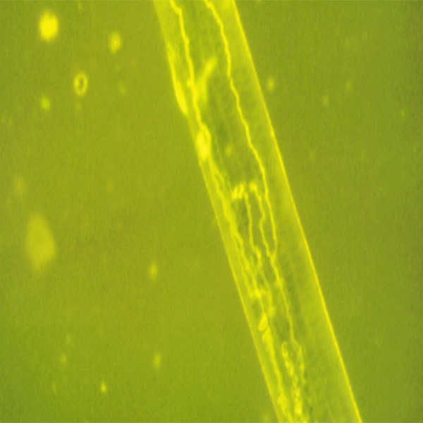
The first step in diagnosing microsporosis is spotting changes in the animal's hair. Such changes are not always present, so veterinarians use Wood's light under which infected hair fluoresces yellow-green.
The most certain diagnosis, however, is from the mycological examination, which involves microscopic and culture examination. Microscopic examination is based on the examination of hair samples and scrapes from the edges of altered parts of the skin, while the culture examination is the cultivation of dermatophytes on an artificial nutrient medium to identify them.


What is Microsporum canis?
Dermatophyte or multicellular fungus Microsporum canis is the most important and common pathogen of dermatophytosis known as microsporosis. It occurs in different animals. This dermatophyte has zoonotic potential, which means that people, most often children, can become infected with it.
How does infection occur?
Stray cats have the most important role in spreading disease. It is essential to know that they do not need to have pronounced signs of illness, while in sick dogs they are almost always present.
How to recognize microsporosis?
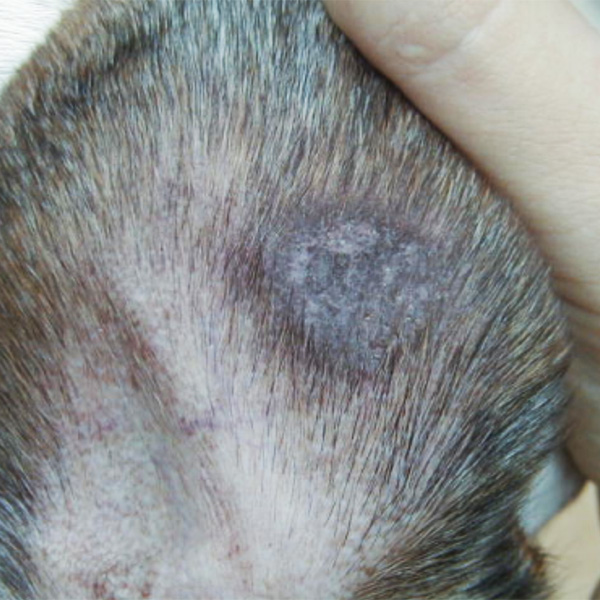
The diseased animal may have thinned out hair at the site of infection, or there can exist oval to round bald areas that are often covered with partially peeled epithelium. This disease is medically called alopecia
How to prevent and how to treat?
Thorough mechanical cleaning and disinfection of the environment in which the animal resides are indispensable steps in preventing the spread of dermatophytoses. An even better effect is achieved by disinfection combined with treatment. Antimycotics are used for general treatment. The treatment is successful and safe, but it must be long lasting for the animal to completely heal.
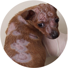

Dog with microsporosis before and after treatment.


The site was co-financed by the European Union, from the European Social Fund.